Sunday, November 27, 2005

Report id’s medico-ethical issues restrict gene therapy growth

Increasing knowledge about how to manage the risks of gene therapy is set to usher in a new era in disease therapies, with the prospect of developing treatments in treating rare, inherited, or life-threatening diseases very real.

Gene therapy has gone to great pains to deal with the toxicological complications and poor therapy efficacy that have hampered the growth of this sector.

So much so that the US gene therapies market, reveals that revenues expect to reach approximately $125 million (€106 million) in 2006 growing to approximately $6541 million in 2011.

This is not to say that gene therapy has not seen its fair share of clinical failures. Poorly regulated trials, and high levels of clinical attrition have seen the promise of gene therapy suffer from negative coverage and unsubstantiated claims.

Despite these remarkable strides, the fact remains that most biotech companies are still years away from having an approved and marketed product.

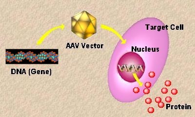
The only exception to this is the China-based company Shenzhen SiBiono GeneTech, whose product Gendicine, made news in October 2003 by becoming the first gene therapy in the world approved for marketing.

The treatment consists of an adenovirus designed to insert a gene called p53. This gene codes for a protein that triggers cell suicide when cells start to run amok, preventing them becoming cancerous.

Many tumours arise after the mutation or inactivation of p53, and in cancers of this type restoring the protein should kill the tumour cells.

The approval signals a breakthrough in the treatment of head and neck squamous cell carcinoma (HNSCC), while ongoing clinical trials expect to uncover more target cancer indications for Gendicine.

China's progress in the cell therapy market has not gone unnoticed by the west, in particular the United States. However, one could argue that US legislation and ethical wrangling concerning stem cells, a crucial factor in cell treatment, has slowed down progress, losing significant ground over rival countries.

"This is critical not only for treating the disease indication and offering hope to patients, but also to validate the whole gene therapy sector to the market and investors," said Raghunath Tantry, Frost & Sullivan's research analyst

"Companies such as Introgen Therapeutics have potentially successful late-stage pipeline products and this will add to the market momentum and increase the credibility of the technology," he added.

Introgen's INGN 241 (breast cancer) is in Phase 1-2 clinical development as is INGN 225 (lung cancer), a therapeutic vaccine, is in Phase 1-2 clinical development.

Also in Phase 1-2 clinical development are INGN 234 (a topical formulation for oral cancer prevention and treatment of oral pre-malignancies), a topical formulation for oral cancer prevention and treatment of oral pre-malignancies and INGN 401 a systemic nanoparticle tumour suppressor.

It seems that pressing concerns such as the medico-ethical issues is likely to have more of an impact on the market's fortunes than any health and safety concerns. Monitoring agencies have even questioned the ethics of testing these therapies on humans, since the science of genetics is still at a nascent stage of development.

"Successes, even minor safety and efficacy milestones, must be communicated to overcome the unwanted publicity received for toxicity and fatalities," commented Tantry.

"The market must be educated on the subtle ways to evaluate a gene therapy product for its benefits to patients that need those therapies."

By Wai Lang Chu

source: drugresearch

Saturday, November 26, 2005

Earliest Animals Had Human-like Genes


image source: http://www.hhmi.org/annual96/research/pix/chart.gif
Species evolve at very different rates, and the evolutionary line that produced humans seems to be among the slowest. The result, according to a new study by scientists at the European Molecular Biology Laboratory [EMBL], is that our species has retained characteristics of a very ancient ancestor that have been lost in more quickly-evolving animals. This overturns a commonly-held view of the nature of genes in the first animals. The work appears in the current issue of the journal Science.

Genes hold the recipes for proteins. The genes of animals usually contain extra bits of DNA sequence, called introns - information which has to be removed as cells create new molecules. The number of introns in genes, however, varies greatly among animals. While humans have many introns in their genes, common animal models such as flies have fewer. From an evolutionary perspective, it was long assumed that the simpler fly genes would be more ancient. The current study reveals the opposite: early animals already had a lot of introns, and quickly-evolving species like insects have lost most of them.

To discover what early animals were like, scientists usually compare their descendents. This is difficult when comparing distantly-related animals such as humans and flies. In these cases, it helps to look at living organisms that have preserved many features of their ancestors. Detlev Arendt's group is doing this with a small marine worm called Platynereis dumerlii. "Similar animals are already found in the earliest fossils from the Cambrium, about 600 million years ago," Arendt explains, "arguing that Platynereis could be something like a 'living fossil'." This makes it an ideal model for evolutionary comparisons to find out what the common ancestors of humans, flies and worms were like."

Until quite recently, such comparisons could only be made by looking at physical characteristics such as the structure of bones, teeth, or tissues. But DNA sequencing now permits scientists to make comparisons of the genetic code and read evolutionary history from it. An international consortium involving researchers from EMBL, the UK, France and the United States has now sequenced a part of the Platynereis genome. "The fraction of Platynereis genes we have been able to look at tells a very clear story," says researcher Florian Raible, who performed most of the computer analyses. "The worm's genes are very similar to human genes. That's a much different picture than we've seen from the quickly-evolving species that have been studied so far."


Raible is member of both Arendt's group and a second EMBL lab, that of Peer Bork, whose specialty is analyzing genomes by computer. "Human genes are typically more complex than those of flies," explains Bork. "Classicallystudied species like flies have far fewer introns, so many scientists have believed that genes have become more complex over the course of evolution. There have already been speculations that this may not be true, but proof was missing. Now we have direct evidence that genes were already quite complex in the first animals, and many invertebrates have reduced part of this complexity."

Not only are the introns there - the team also discovered that their positions within genes have been preserved over the last half a billion years." This gives us two independent measurements that tell the same story," Raible explains. "Most introns are very old, and they haven't changed very much in slowly-evolving branches of life, such as vertebrates or annelid worms. This makes vertebrates into something like 'living fossils' in their own right."

The discovery that Platynereis also represents a slowlyevolving branch of animal life has important implications for the study of humans. "We've already learned an incredible amount about humans from studies of the fly," Arendt says. "The marine worm might well give us an even better look at important conserved processes. Another thing that this has shown us is that evolution is not always about gain; the loss of complexity can equally be an important player in evolution."

Source article:

Vertebrate-type intron-rich genes in the marine annelid Platynereis dumerilii
F. Raible, K. Tessmar-Raible, K. Osoegawa, P. Wincker, C. Jubin, G. Balavoine, D. Ferrier, V. Benes, P. de Jong, J. Weissenbach, P. Bork and D. Arendt.
Science, 25 November 2005

The original news release can be found here.

Friday, November 25, 2005

Clone human embryo creator admits ethical lapses in work

Alok Jha, science correspondent
Friday November 25, 2005

The scientist who created the first clone human embryo was forced to apologise yesterday for ethical lapses in his work. Professor Woo-Suk Hwang of Seoul National University said some of the eggs used in his experiments to create human clones were donated by two junior members of his research team. He admitted his focus on the research had led him to overlook the related ethical issues.

An investigation by the South Korean health ministry also found some women were paid for their eggs, a practice that is now illegal in the country. "I am very sorry that I have to tell the public words that are too shameful and horrible," said Prof Hwang at a news conference yesterday. "I should be here reporting the successful results of our research, but I'm sorry instead to have to apologise."

Prof Hwang said he would resign as the head of the World Stem Cell Hub in a bid to "atone to the public". The hub, launched in Seoul last month, aims to help coordinate global efforts to use stem cells in the search for treatments for incurable diseases such as Alzheimer's and Parkinson's. The group had announced plans to open laboratories in San Francisco and England, but Prof Hwang's close collaborators, including University of Pittsburgh researcher Gerald Schatten, dropped out of the project following the allegations of unethical egg collection.

A hero in his native South Korea, Prof Hwang made headlines last year when he announced he had extracted stem cells, the master cells that can turn into any type of cell, from the first cloned human embryo. Earlier this year he unveiled Snuppy, the world's first cloned dog.

Reports that Prof Hwang may have used eggs from members of his research team first appeared in the scientific journal Nature last year. Prof Hwang said this was the first time the issue had been brought to his attention and that when he asked the scientist in his team, she admitted donating eggs but asked for her privacy to be maintained.

Prof Hwang said: "I have learned a painful lesson that I should conduct research in a calm and cautious manner by living up to a global standard."

But Prof Hwang's Seoul team are unfazed by the revelations. Lee Jeong-Ryul, a cardiac surgeon, said the dispute was "no reason to stop stem cell research".

source: The Guardian

Thursday, November 24, 2005

Hair follicle stem cells repair nerve damage: study

Los Angeles | November 23, 2005 11:15:06 AM IST

Patients with an injured nervous system could be cured with their own hair follicle in future, as US and Japanese scientists have succeeded in curing severe nerve damage in mice by using tissues differentiated from hair follicle stem cells.

The results of the new research suggest hair follicle stem cells can promote nervous axon growth and functional recovery after nerve injury, thus offering an opportunity for the clinical treatment of peripheral nerve diseases, said the researchers.

The team, including researchers from the Massachusetts Institute of Technology, the Kitasato University of Japan, and the University of California, San Diego, reported this achievement in the latest issue of the journal the Proceedings of the National Academy of Sciences.

Embryonic stem cells, known to be capable of differentiating into almost all tissue cells, have aroused ethical debates in many countries. Scientists also found that problems such as immunologic incompatibility are linked with embryonic stem cells.

Therefore, more recent studies have focused on adult stem cells for future clinical applications. And hair follicles afford a highly promising source of relatively abundant and accessible, active, pluripotent adult stem cells, said the researchers.

In earlier studies, the team led by Robert Hoffman, a professor at the University of California, San Diego, has induced hair follicle stem cells to differentiate into blood vessel cells and neurons. The researchers said these studies suggested the potential of hair follicle stem cells to form diverse cell types.

Now the researchers have successfully coaxed the hair follicle stem cells to evolve into the Shwann cells, a variety of glia cells that wrap around axons in the peripheral nervous system.

When injected into disabled mice with injured sciatic nerve, these Shwann cells produced myelin sheaths that surround nerve axons, and then the mice were able to walk normally, the researchers reported.

"Therefore, by differentiating into Schwann cells, the hair follicle stem cells may stimulate the host axons to extend and, thus, to fill the transaction gap," they said in the paper.

Hair follicle stem cells may be more promising in therapies, according to the researchers. In the future, patients with injured nervous system could be cured with their own hair follicle.

--Xinhua


(IANS)

Wednesday, November 16, 2005

Vietnamese scientists decode gene of bird flu virus


H5N1 Virus
Vietnamese scientists have decoded the gene of H5N1 virus, paving the way for defining the variations and transmission mechanism of the bird flu virus, local newspaper Youth reported Wednesday.
Vietnam's Ho Chi Minh City Pasteur Institute and the Regional Veterinary Centre in the southern city claimed Tuesday they have entirely decoded the gene of the virus.

Ngo Bao Long, of the Veterinary Centre, said they found not only the virus strain H5, but also two other strains of H3 and H4 in two samples from poultry.

Theoretically, when a fowl is infected with H5, H3 and H4 at the same time, the viruses can swap their genes to create a new virus strain, which can be more dangerous, he said.

Vietnam, in early 2006, is likely to churn out 20-50 million dozes of H5N1 vaccines to be used for poultry next year, said the biotechnology institute's director Le Tran Binh, adding his institute has completed procedures to produce the vaccines.

Meanwhile, the country's National Institute of Hygiene and Epidemiology is completing final procedures to produce H5N1 vaccines to be used for people. The Hanoi-based institute that has been involved in research into the vaccines since 2004, has proposed the health ministry use them on trial basis in early 2006.

Since December 2004, Vietnam has detected 65 human cases of bird flu infections, including 22 fatalities, according to the health ministry.

--Xinhua

source: (IANS)


Friday, November 11, 2005

Heart risk gene hits African Americans hardest

A gene commonly found in Americans of European descent can be deadly when carried by African Americans, a new study has revealed. The gene variant more than triples the risk heart attack in African American populations, the researchers found.

African Americans are known to be more prone to heart attacks and the researchers suggest this may partly be due to European ancestry in those individuals, although environmental factors are certainly involved.

Kári Stefansson at Decode Genetics in Reykjavik, Iceland, and colleagues, isolated a gene variant called HapK, which is found in 30% of European Americans and 6% of African Americans. The variant also occurs in about 35% of people in Asia, but native Africans do not possess the gene. Stefansson proposes that the gene mutation must have occurred after the migration of human populations from Africa, about 50,000 years ago.

HapK is involved in series of biochemical steps that leads to inflammation in the body. Its role is not fully understood but it is believed to increase the propensity of fatty deposits in the arteries – atherosclerotic plaques – to rupture, leading to a heart attack.

250% increased risk
Stefansson studied the significance of the gene variant in more than 3000 people in three groups, one at the Cleveland Clinic in Ohio, one at Emory University in Atlanta, and one at the University of Pennsylvania in Philadelphia, all in the US.

In the study, published in Nature Genetics, they found that HapK was associated with a slight increase in risk of heart attack for participants of European descent – just 16% – while those of African descent were 3.5 times more likely to suffer a heart attack (equivalent to a 250% increase in risk).

“If you’re an African American with the variant gene you are close to certain to have a heart attack if nothing is done about it,”"It’s very important to screen and find this subgroup,” Stefansson says. His biopharmaceutical company has two potential drug therapies in clinical trials which act by regulating the inflammatory pathway that HapK is involved in.

Micro-flora defence
Martin Godfrey, a physician at the British Cardiac Society, notes: “There are significant ethnic variations in heart attack risk. Black people in Britain and America are particularly at risk and they have increased incidence of strokes and high blood pressure.”

Stefansson speculates that the variant is so dangerous in African Americans because, unlike European Americans, they have not had thousands of years to adjust to its presence in their genome.

“The inflammatory pathway probably developed as a protective response to micro-organisms; and as populations inhabited very different areas, the micro-flora they encountered was vastly different,” he says. “That may be the reason for the differences in frequency of the variant.”

Journal reference: Nature Genetics

source: morover

Thursday, November 10, 2005

Some 32,000 genes to go in squid research project

WOODS HOLE, Massachusetts The room is filled with marine life: fiddler crabs and moon snails, dogfish and flounder. There are clams stacked up by the dozen, with signs that require second looks. "Toadfish infected with lice," one sign says. "Do not touch."

But Joe DeGiorgis, 41, walks right past it all. He has come to the Marine Resource Center in Woods Hole for one thing and one thing only.

"The squid are here," he calls out, pointing to a large oval tank. "My favorite guys."

DeGiorgis, smiling, peers inside the tank. It's filled with dozens of long-finned Atlantic squid - 12 to 18 inches in length, or about 30 to 46 centimeters long, typical in every way as far as squid are concerned.

DeGiorgis likes fried calamari as much as the next guy. But as a post-doctoral fellow at the National Institutes of Health who spends half the year working at the Marine Biological Laboratory in Woods Hole, he has other reasons for admiring these mollusks.

He and a colleague, J. Peter Burbach, a professor of molecular neuroscience at University Medical Center Utrecht in the Netherlands, are the minds behind the Squid Genome Project, a surprisingly successful attempt to map the squid's genetic thumbprint in the hopes that its secrets may help solve mysteries like Alzheimer's disease.

On a budget of roughly $100,000, DeGiorgis and Burbach have identified more than 3,000 of the squid's estimated 35,000 genes, including, DeGiorgis said, the gene that causes the body to produce insulin, and genes linked to

Alzheimer's and a degenerative and fatal neurological disease called Niemann-Pick Type C in humans.

That's no small accomplishment. Stephen Sturley, an assistant professor of pediatrics at Columbia University, said DeGiorgis's work may one day help doctors find a cure for Niemann-Pick. And that, says DeGiorgis, is only the beginning.

"If the project were to stop today," he says, "I've got enough genes to work on for the rest of my career."

DeGiorgis grew up on Jacques Cousteau television shows and took up scuba diving in high school. He got his first job at the Marine Biological Laboratory in Woods Hole before his senior year in college.

It wasn't much, just a diving job. DeGiorgis dove for surf clams in the sandbars off Martha's Vineyard and brought them back to Woods Hole for scientists to study. He came to learn that the scientists were interested in what marine life, namely clams and squid, could reveal about people.

Squid, he learned, are especially powerful models. Their giant axon, a channel of nerve fibers in their back, is 1,000 times wider than the average human axon - an easy target for researchers who want to study how information travels and systems break down. DeGiorgis set up a private company to do dissections of the axon and other oddities. For a time in Woods Hole, he was the guy you called when you needed, say, 2,000 squid eyes, dissected and ready to be studied.

As DeGiorgis got his doctorate in cell and molecular biology from Brown University in 2001, he began to focus on mapping the squid genome.


"Some people were like: 'What would you do with it if you had it?' They didn't understand the significance of the work," he says. But DeGiorgis knew the squid could provide insight into the human condition.

DeGiorgis peers down at the squid in the water. He nets one, and it promptly sprays him in the face with black ink. DeGiorgis smiles.

Squid, he says, are beautiful.

source: IHT

Wednesday, November 09, 2005

Researchers Discover New Form Of Cancer Gene Regulation

Now, a team of researchers from Northwestern University and the University of Wisconsin has shown that the Quaking gene likely suppresses tumor growth by inhibiting production of a protein associated with GLI1, a cancer-causing oncogene highly associated with severe birth defects and several childhood cancers.

The group's study, published in the Nov. 1 online issue of Developmental Biology, details the discovery of an important and completely novel form of regulation of the GLI1 gene.

"Results of the study open a new research direction for issues ranging from cancer formation to environmental interactions in development and will point the way to similar mechanisms of control in other genes," said Philip M. Iannaccone, M.D., who led the study.

Iannaccone is George M. Eisenberg Professor of Pediatrics at Northwestern University Feinberg School of Medicine and deputy director for basic research at Children's Memorial Research Center.

Development occurs as a coordinated series of genetic control events that create proliferation of cells, signals for further differentiation, proteins that define cellular function and "programmed" movement of cells into developing structures.

These processes, known as pattern formation, are controlled largely by networks of genes and proteins called signal transduction pathways that receive signals from outside of the cell in the form of protein interaction with the cell surface. Through a series of intracellular events, these signals trigger gene activation or repression through action of transcription factors in the nucleus of the cell.

The altered gene expression profile then results in cellular differentiation, cellular proliferation or cellular death as pattern formation proceeds.

An important signal transduction pathway critical to early development of humans and animals involves the genes Sonic hedgehog (the signal) and GLI (the transcription factor).

The GLI family of three genes was first discovered in a human brain tumor, and mutations in this family of genes result in severe birth defects and devastating cancers in humans.

"While some cancers are explained by known defects in the regulation of the GLI1 gene, for many cancers the reasons for excessive GLI1 protein are not known. The protein levels and activity of GLI1 are likely regulated at levels other than the gene," Iannaccone said.

The form of regulation the researchers discovered occurs after the gene makes messenger RNA, the first step toward making a protein that controls cell fate. Once the messenger RNA leaves the cell, it participates in a process called translation, during which the cellular machinery makes a protein by linking amino acids together according to the plan described in the messenger RNA and thereby based on the information from the DNA sequence of the gene.

Iannaccone and colleagues showed that after the messenger RNA for GLI1 is made, it binds to the Quaking protein and inhibits the translation event. This means that all of the controls that the cell has on the gene for GLI1 can be present and active and the GLI1 is still not produced.

Significantly, the study demonstrated that this regulation is conserved from human to the worm, Caenorhabditis elegans (often used in laboratory research), indicating that the formation of these RNA protein complexes is a very ancient form of regulation of protein function.

Olga Lakiza, postdoctoral fellow in pediatrics at the Feinberg School, was the first author on the article. Iannaccone's other co-researchers on this study included David O. Walterhouse, associate professor of pediatrics, Feinberg School and Children's Memorial Research Center, and Elizabeth B. Goodwin, department of genetics, University of Wisconsin, Madison.

source: sciencedaily

Scientists discover anti-ageing gene

Scientists in the United States have discovered that a gene prolonging life in mice works by controlling insulin.

The gene, Klotho, is found in several species. In mice, the researchers discovered, it acts as a hormone, circulating through the blood and binding to cells.

"Therapies based on this hormone could prove to be a way to extend life or slow its effects," Dr Makoto Kuro-o, assistant professor of pathology at the University of Texas' Southwestern Medical Center and senior author of the study, told Science magazine.

"It could be one of the significant steps for developing anti-aging therapy."

Dr. Kuro-o and his colleagues originally discovered the Klotho gene in 1997, naming it after one of the mythical Greek fates who controlled the length of human life. Their previous studies have shown that mutant mice lacking the Klotho gene appear normal until about 3 to 4 weeks old, and then begin showing signs of age, such as skin atrophy, osteoporosis, arteriosclerosis and emphysema. The mice died prematurely at about two months.

For the current study, they created a second strain of mutant mice in which the Klotho gene generated more of the protein than in normal mice. Those mice lived between 19 per cent to 31 per cent longer than normal mice.

The researchers were especially interested in how the hormone affected insulin, because making an animal resistant to insulin increases its lifespan - a phenomenon found in animals ranging from worms to fruit flies to mice.

The mice with higher levels of Klotho had more insulin in their system than the normal mice, suggesting that the Klotho mice were resistant to insulin. The opposite was true with the mice deficient in Klotho. They were more sensitive to insulin and had reduced levels of it.

But there may be downsides with Klotho.

The long-lived mice in the new experiments had fewer offspring than normal mice.

And the gene may also predispose people to diabetes.

source:999today

Super-fast robot muscles proposed

A new theory has been developed for creating robot muscles that are 1,000 times faster than human muscles.

The muscles would have a simpler design than existing robot muscles and have no additional energy demands.

Current robotic muscles move 100 times slower than human muscles.

MIT researchers led by Professor Sidney Yip have proposed a new theory that might eliminate one obstacle to speeding them up.

An MIT news release reports:

In this case, a robotic muscle refers to a device that can be activated to perform a task, like a sprinkler activated by pulling a fire alarm lever, explains Yip, a professor of nuclear engineering and materials science and engineering.

In the past few years, engineers have made the artificial muscles that actuate, or drive, robotic devices from conjugated polymers. "Conjugated polymers are also called conducting polymers because they can carry an electric current, just like a metal wire," says Xi Lin, a postdoctoral associate in Yip's lab. (Conventional polymers like rubber and plastic are insulators and do not conduct electricity.)

Conjugated polymers can actuate on command if charges can be sent to specific locations in the polymer chain in the form of "solitons" (charge density waves). A soliton, short for solitary wave, is "like an ocean wave that can travel long distances without breaking up," Yip adds. (See figures.) Solitons are highly mobile charge carriers that exist because of the special nature (the one-dimensional chain character) of the polymer.

Scientists already knew that solitons enabled the conducting polymers to conduct electricity. Lin's work attempts to explain how these materials can activate devices. This study is useful because until now, scientists, hampered by not knowing the mechanism, have been making conducting polymers in a roundabout way, by bathing (doping) the materials with ions that expand the volume of the polymer. That expansion was thought to give the polymers their strength, but it also makes them heavy and slow.

Lin discovered that adding the ions is unnecessary, because theoretically, shining a light of a particular frequency on the conducting polymer can activate the soliton. Without the extra weight of the added ions, the polymers could bend and flex much more quickly. And that rapid-fire motion gives rise to the high-speed actuation, that is, the ability to activate a device.

To arrive at these conclusions, Lin worked from fundamental principles to understand the physical mechanisms governing conjugated polymers, rather than using experimental data to develop hypotheses about how they worked. He started with Schrödinger's equation, a hallmark of quantum mechanics that describes how a single electron behaves (its wave function). But solving the problem of how a long chain of electrons behaves was another matter, requiring long and complex analyses.

source: betterhuman

Gene silencing and microRNA discovery


Biogenesis and function of microRNA
Medical Science News

One of the body's primary strategies for regulating its genome is a kind of targeted gene silencing orchestrated by small molecules called microRNAs, or miRNAs.
First observed only a few years ago, these molecules appear to inactivate messenger RNA, itself responsible for translating genes into proteins. Scientists have been eager to know more about miRNAs, clearly important players on the genetic field despite having gone unnoticed for so long. How are they produced? And how do they work?

In a series of studies published over the past year, a research team at The Wistar Institute has provided considerable insight into the world of miRNAs. In their first study, which appeared last year in Nature, they identified a two-protein complex, called the microprocessor, which controls the earliest steps in the creation of miRNAs in the cell nucleus.

In their next study, published in Nature earlier this year, the Wistar group described a three-protein complex that picks up the process in the cell cytoplasm and carries it through to the maturation of the finished miRNAs.

Now, in new findings published online November 3 in Cell, Wistar professor Ramin Shiekhattar, Ph.D., and his colleagues report that the three-protein complex has been identified as RISC, a previously glimpsed but ill-defined molecular complex known to be involved in gene silencing. RISC, the new study demonstrates, not only oversees production of miRNAs, as described in the earlier study this year, but is also responsible for miRNA specificity in silencing particular messenger RNAs.

In RISC, two of the three components, Dicer and Argonaute 2, are enzymes bound together by the third member, TRBP. Dicer cuts double-stranded precursor molecules shaped like hairpins into pairs of short single-stranded miRNAs - in essence, nipping off the bend in the pin. RISC then unzips the two single-stranded miRNAs from each other and identifies and holds one as a guide to help it find the specific messenger RNA to be inactivated. Using complementarity to match the guide miRNA to a particular length of its target messenger RNA, Dicer and TRBP then hand over the messenger RNA for cutting by Argonaute 2. Still holding the guide miRNA, RISC then scouts for additional copies of its target messenger RNA to cut. The cutting destroys the messenger RNA, effectively silencing the gene from which it was transcribed.

"The two enzymes in the complex are like two scissors working together in a concerted fashion, connected and coordinated by the third member of the complex," Shiekhattar explains.

Another scientific question surrounding RISC was also resolved by the current study. Some investigators had theorized that the activity of RISC required ATP for energy. ATP, or adenosine triphosphate, is a molecule used to store and release energy for tasks throughout the body.

"The work of RISC is being accomplished with no energy requirement whatsoever," Shiekhattar says. "All of the activity - the separation of the strands, the multiple cutting steps, everything - is being done in the absence of any energy use."

Instead, he says, the different molecules involved have stronger and weaker affinities for each other that govern their stepwise associations and disassociations as the process unfolds.

http://www.wistar.upenn.edu/

Study shows link between gene mutation and efficacy of cancer drugs


Human melanoma cell undergoing cell division.
A team of researchers led by scientists at the Memorial Sloan-Kettering Cancer Center have discovered that a new class of drugs, now in early stage clinical trials, works best in patients with mutations in the BRAF gene.

BRAF is a protein that plays a central role in the growth and survival of cancer cells and is mutated in the majority of patients with melanoma and in a minority of patients with colon, breast, and lung cancers. The findings represent a potential targeted therapy tailored for patients whose tumors contain this mutation.

The researchers found that drugs that inhibit a protein called MEK selectively inhibited the growth of cancer cells lines and tumors that have a mutated BRAF gene. One of these drugs, PD0325901, developed by Pfizer, is now being tested in clinical trials of patients with melanoma, colon, breast, and lung cancers.

In addition, by re-analyzing the data on more than 42,000 compounds tested by the National Cancer Institute against a panel of 60 cancer cell lines, the investigators were able to identify a small number of other compounds that also selectively inhibit tumors that have the BRAF mutation.

While the mechanism of action of some of these compounds has yet to be determined, several of the most effective compounds were also inhibitors of the MEK protein.

"We find that all tumors with the BRAF mutation and some with the RAS mutation are sensitive to drugs that inhibit MEK," explained Dr Neal Rosen, laboratory head in the Molecular Pharmacology and Chemistry Program at Memorial Sloan-Kettering and the study's senior author. "Translating these findings into a strategy for treating patients whose tumors are dependent upon this specific genetic change is the next step, and such clinical trials are now ongoing."

source: pharmabusiness

Tuesday, November 08, 2005

Gene doping is a real danger - but anti-doping science is making progress

Gene therapy research has reached a critical phase. Already practised on humans as part of strictly controlled experiments, gene therapy promises to become a widely available form of treatment for injury and disease.

However, advances in the science of gene therapy have a darker side: gene doping - the unscrupulous use of genetic modification to enhance athletic ability by athletes, sportspeople and coaches.

"We have seen an interest among individuals who contact gene researchers for the purpose of doping in sport," said Karolinska Institutet's Professor Arne Ljungqvist, Sweden's most well-known anti-doping expert and chairman of WADA's (World Anti-Doping Agency) Health, Medical and Research Committee. "This is a disturbing trend because not only is gene doping in sport wrong, it can also be extremely dangerous."

The current status of research in the field of gene-doping detection will be presented at an international symposium to be held at Karolinska Institutet in Stockholm, Sweden on 4 - 5 December 2005. Two press conferences will be held in connection with the symposium (see below), which will be attended by the world's leading gene researchers and some of the sporting world's most prominent personalities.

"Gene doping represents a serious threat to the integrity of sport and the health of athletes," said WADA chairman Richard W. Pound. "As the international organisation responsible for promoting, coordinating and monitoring the global fight against doping in sport in all its forms, WADA is devoting significant resources and attention to ways that will enable us to detect gene doping so that we can catch the cheaters, level the playing field and ensure the safety of athletes. The 2nd WADA Symposium on Gene Doping promises to help advance these efforts."

One of the most important messages of the symposium is that anti-doping scientists are working vigorously alongside genetic scientists so that, as new therapeutic methods are being developed, anti-doping scientists are finding new ways to detect gene doping.

"Gene doping will in all likelihood soon be with us, and I would not be surprised if the first tentative steps had already been taken," said American professor Theodore Friedman, one of the world's leading gene researchers, chairman of WADA's Gene Doping Panel and the first speaker at the symposium.

Sportspeople are taking immense risks when they add new genetic material into their bodies. Already there have been at least two deaths during experiments conducted to treat the sick.

"Two people have, for example, developed leukaemia," continued Professor Friedman. "The seriously ill can take such a risk perhaps, but for young, healthy sportsmen and women, it is completely unacceptable."

One challenge that anti-doping experts are trying to tackle is the fact that gene therapy methods, once available, will be relatively simple to use. All that may be needed is a standard laboratory.

The genes attractive to sport are well defined: those that stimulate tissue growth and boost strength, and those that increase stamina by stimulating the production of red blood cells.

On 4-5 December 2005, Stockholm is hosting the 2nd WADA SYMPOSIUM ON GENE DOPING. Participants include some fifty leading research scientist in the field of gene technology and gene therapy from around the world. The symposium is being organized by the WADA (World Anti-Doping Agency) in association with the Swedish Sports Confederation and Karolinska Institutet, and is a follow-up to the first WADA gene doping symposium held in New York's Banbury Centre in March 2002.

source: medicalnews

Tokyo Nanotech 2006

The Micro and Nanotecnology (MNT) Network is looking for 20 companies in the nanotech sector to attend Tokyo Nanotech 2006, which takes place 21-23 February next year.

This is an excellent opportunity for companies to showcase their nanotechnology offering through subsidised exhibition space at a major industry event. Tokyo Nanotech 2005 was very successful in raising the profile of UK excellence in nanotechnology and the 22 exhibiting companies enjoyed good business development opportunities leading to 589 qualified leads. The exhibition in 2006 will have larger floorspace and a more focused presentation to ensure maximum benefit to participants.

EEDA has been asked to submit nominations from the East of England. A shortlist of nominations must be submitted to MNT Network by 18 November. In order to allow time to evaluate nominations from the region please respond to EEDA by Monday 14 November.

Potential exhibitors should :

Explain how they see their significance in UK and global terms
show how their work leads into beneficial applications
explain how they would benefit from exhibiting at Tokyo Nanotech 2006 show how their work could be presented visually and by a team give background details of the company structure, history, personnel Agencies or consultancies cannot apply - you must have a strong product or process offering.

Showcase 'virtual exhibitors'

Additionally, we would like to receive bids from companies with compelling technology demonstrations, whether they be materials, processes, video based content, or pieces of equipment. These will compete for space in the showcase area, where MNT Network is planning a 'science-museum' type self-navigated experience. Companies applying for this presence would not be required to attend the event in person, but must be prepared to organise the shipping of equipment and supply information so that explanatory graphics can be produced.

For the selected companies all exhibition costs will be covered; companies need only cover the travel and subsistence costs of their representatives.

If you would like to be considered for nomination, please send your submission by Monday 14 November to:

Adam Bryan (adambryan@eeda.org.uk), tel: 01223 202807

Nanotech breakthrough aids quest for viable alternative energy sources

T
A photovoltaic solar cell
At a time when oil prices are reaching record highs and people are bracing for winter heating bills, researchers at Wake Forest University's Center for Nanotechnology and Molecular Materials have made significant strides in improving the efficiency of organic or flexible solar cells.

Traditional silicon solar panels are heavy and bulky and convert about 20 percent of the light that hits them to useful electrical power. For years, researchers have worked to create flexible, or "conformal," organic solar cells that can be wrapped around surfaces, rolled up or even painted onto structures, but the best scientists have been able to do is about 3 percent efficiency, until now.

Researchers at Wake Forest, with the help of researchers at New Mexico State University, have achieved an efficiency rate for organic solar cells of almost 6 percent. In order to be considered a viable technology, the solar cells must be able to convert about 10 percent of the energy in sunlight to electricity. Wake Forest researchers hope to reach 10 percent by October 2006, said David Carroll, director of the nanotechnology center at Wake Forest.

"The consumer market would be really open to having these conformal systems if you could, for instance, roll them up and put them away," said Carroll, who is also an associate professor in Wake Forest's physics department. "Imagine a group of hikers with a tent that when you unrolled the tent and put it up, it could generate its own power. Imagine if the paint on your car that is getting hot in the sun was instead converting part of that heat to recharge your battery."

Carroll said flexible, organic solar cells also offer several possibilities for military use.

"The military would obviously want something like that because you could only put maybe tens of those big solar panels on a transport, but you could put hundreds of ultra-thin flexible ones on a transport and supply half the army," he said.

Most experts have estimated that flexible, solar cell technology for consumers was about a decade away, but Carroll said the new breakthrough at Wake Forest and NMSU means that consumers could be using this technology in the next five years.

Using a set of polymer coatings, researchers at Wake Forest constructed a nanophase within the polymer called a "mesostructure." The "mesostructure" changes the properties of the plastic and makes it better for collecting light. The researchers also removed the current from the polymer coating, Carroll said.

A test system at Wake Forest's nanotechnology center was used to simulate the sun, Carroll said, and the simulated spectrum was precisely measured and shot onto the organic solar cell, which appeared as a thin coat of paint. Devices at the center have registered almost 6 percent efficiency.

This breakthrough was announced in October at the Santa Fe Workshop on Nanoengineered Materials and Macro-Molecular Technologies, which was sponsored by Wake Forest's nanotechnology center.

Source: Wake Forest University (By Jacob McConnico)

Monday, November 07, 2005

Gene Therapy in a Bottle of Mouthwash

Brush your teeth and rinse with gene therapy. That could be the future of oral health care as envisioned by Colgate-Palmolive and Introgen Therapeutics, an Austin, Tex., biotechnology company.

The companies announced an alliance Friday to incorporate gene therapy - an exotic, experimental and so far largely unsuccessful form of medicine - into mouthwashes, gels and similar products to treat and prevent oral cancers.

Colgate is buying about 3.6 million shares of Introgen, or a 9.7 percent stake. Colgate will pay $20 million, or $5.54 a share, the same as Introgen's closing price on Thursday. On Friday, Introgen shares climbed 8 percent, to $5.96.

Gene therapy involves putting genes into cells in the body, often to replace native genes that are malfunctioning. The technique has rarely worked in the 15 years it has been tried, largely because of the difficulty of getting enough functioning genes into cells.

Introgen's method puts so-called tumor suppressor genes into cancerous cells to stop the growth of tumors. The company's most advanced drug, which is in late-stage clinical trials, is a treatment for head and neck cancer that it hopes will be the first gene therapy approved in the United States. In that treatment, viruses containing the desired gene are injected directly into tumors.

Working with Colgate, Introgen will try to put the tumor suppressor genes into an oral product to treat leukoplakia, a precancerous condition characterized by lesions on the cheeks, gums or tongue. In some cases the lesions turn cancerous, usually after many years. Dr. Robert E. Sobol, Introgen's senior vice president for medical and scientific affairs, said an initial product would probably be a prescription drug that would require approval by the Food and Drug Administration. But he said he hoped eventually the product could be sold over the counter.

The company is already in early stages of human testing of a mouthwash containing a tumor-suppressor gene. The mouth is "just such an easy place to apply these therapies," said Dr. Sobol, explaining that it was easy to reach and see the damaged cells. Nevertheless, getting enough genes into cells in the mouth could still be difficult.

Introgen puts its gene into a disarmed cold virus, called an adenovirus, which infects cells and carries in the gene. While Dr. Sobol said the technique was safe, an adenovirus was used in a gene therapy experiment that killed a teenager at the University of Pennsylvania in 1999. Dr. Sobol said that if Introgen wanted to develop a product that would be used widely by people who do not yet have cancer, it might have to use a gene therapy approach that did not involve a virus.

Colgate, which is based in New York, said in a statement that the alliance would give it "insight into a very exciting technology with potential application to oral health."

The company said the alliance was not a move into the pharmaceutical business but rather one of many research efforts it has involving oral health. Known mostly for consumer products, Colgate sells products used by or prescribed by dentists, including the PerioGard antibiotic rinse to fight the gum disease gingivitis.

source: NYT

Next-generation DNA microarray manufacturing

New microarray printing technology is currently producing single-array microarrays with 95,000 and 185,000 features on standard 1x3' glass slides and microarrays containing eight arrays per slide

Agilent Technologies has announced a next-generation DNA microarray manufacturing process that increases feature density more than fourfold in 2006 and potentially greater than tenfold in 2007.

This move is part of Agilent's growing push to develop microarray applications that augment traditional gene expression analysis and enable researchers to answer previously intractable biological questions.

The new microarray printing technology, a higher-performance version of Agilent's proprietary ink-jet-based in situ fabrication method, is currently producing single-array microarrays with 95,000 and 185,000 features on standard 1x3' glass slides.

Agilent is also using the new manufacturing process to print microarrays containing eight arrays per slide.

Multiple-array slides containing user-defined content enable scientists to analyse focused gene sets, biological pathways or chromosomal regions at significantly lower cost per array, using a more automated workflow.

Agilent's current single-array microarrays have 44,000 features.

The company's next-generation microarrays, in single- as well as multiple-array slide formats, will be available in the second quarter of 2006.

'Density is important to the ChIP-on-chip work we do,' said Richard Young of the Whitehead Institute.

'In our laboratory the new Agilent microarrays are boosting productivity by letting us cover the same genomic regions faster and with less expense compared with the previous generation.

'The higher feature density complements the Agilent platform's very high sensitivity and signal-to-noise ratio as well as its well-known flexibility of content.

'All of this makes this platform particularly well suited to emerging applications'.

'Agilent's ink-jet manufacturing process produces the highest sensitivity microarrays in the industry and offers unparalleled content flexibility,' said Mike Booth, general manager, Genomics business, Agilent Integrated Biology.

'These two advantages, combined with higher feature densities, will place us in a unique position to deliver both a broad range of new applications as well as lower microarray costs to life science researchers'.

Agilent's higher-density microarrays will be available as standard catalogue products as well as user-designed custom microarrays for a range of genomic applications including gene expression studies, comparative genomic hybridisation (CGH), ChIP-on-chip and splice variant analysis.

The high-density capability will benefit these as well as future applications by providing a more practical path to whole genome experiments.

In addition, by pairing higher density with Agilent's unique ability to ink-jet-print custom microarray content in multiple-array slide formats, the company will enable more automated workflows and significantly reduce the cost of microarray experiments.

This will make high-quality commercial microarrays accessible to customers who previously could not afford them.

The flexibility of Agilent's microarray printing technology is delivered to customers through the company's eArray website.

'Scientists are increasingly exploring multiple applications and want to validate gene expression data with other types of data,' continued Booth.

'Agilent has aggressively augmented our informatics portfolio in recent months to facilitate this multi-application trend, and our new, higher-density microarray capability further supports this approach'.

In addition to increased feature density, the enhanced ink-jet printing provides improved spot placement accuracy and further optimised synthesis uniformity.

This enables features to be synthesised closer together than on the previous generations of microarrays with minimal impact on feature size.

The 95,000 and 185,000 feature microarrays are therefore compatible with existing experimental protocols without the need for Agilent customers to make further capital investment in a new scanner.

Agilent is a provider of microarray-based, genomics research systems.

This includes reagents for Sample Preparation and microarray processing, hardware for sample QC and high-throughput microarray scanning, 60-mer oligo microarrays on industry-standard 1x3' glass slides for gene expression, comparative genomic hybridisation and chromatin immunoprecipitation applications, custom microarray design services, and Genespring and Rosetta software products for data analysis.

source: laboratory talk

Sunday, November 06, 2005

Fighting Cancer from the Inside Out

Manmade molecules that deliver drugs directly to cancer cells, tiny sensors that monitor oxygen levels in the bloodstream, molecular surgery to remove defective genes – it all sounds like science fiction. But technology to make these advances possible is being developed today by scientists at the Michigan Nanotechnology Institute for Medicine and the Biological Sciences (M-NIMBS).

“Nanotechnology allows us to make materials that are thousands of times smaller than the smallest cell in the body,” says James R. Baker Jr., M.D., Ruth Dow Doan Professor of Biologic Nanotechnology at the University of Michigan, who directs the Michigan Nanotechnology Institute. “Because these materials are so small, they can easily get inside cells and change how they work.”
So how small is small?

One nanometer equals one-billionth of a meter, which means it would take about 80,000 nanometers lined up side-by-side to equal the diameter of a human hair. “To compare the size of a nanoparticle to the size of a typical cell, think of a grain of sand on a football field,” Baker says.

When it comes to fighting cancer, scientists are excited about developing new nanotechnology-based treatments that will be more effective and have fewer side-effects than traditional therapies. As an example, Baker points to the recent creation, by M-NIMBS scientists, of what he calls the “nanotechnology equivalent of a Trojan horse.”


Dendrimer
It's a manmade nanoparticle called a dendrimer designed to smuggle a powerful anti-cancer drug inside tumor cells – increasing the drug's cancer-killing activity and reducing its toxic side effects. Less than five nanometers in diameter, the particle is small enough to slip through tiny openings in cell membranes.

Dendrimers have a tree-like structure with many branches where scientists can attach drugs and molecules. U-M scientists attached methotrexate, a powerful anticancer drug, to one branch of the dendrimer. On another branch, they attached their secret ingredient – a vitamin called folic acid.

Folic acid, or folate, is an important vitamin required for the healthy functioning of all cells. But cancer cells, in particular, seem to need more than average amounts.

By taking advantage of a cancer cell's appetite for folate, U-M scientists were able to concentrate more of the toxic drug in cancer cells, while reducing side-effects on normal cells.

“It's like a Trojan horse,” Baker explains. “The cancer cell thinks it's bringing in food. Once inside, however, there's a poison on the nanoparticle that kills the cell.

When Baker and his research team gave the nanoparticle-methotrexate combination to mice with tumors, they found it was more effective than giving the cancer-killing drug alone.

“Effectively, we delayed the growth of tumors in mice for 30 days,” Baker says. “That is significant, when you consider that one month for a mouse is the equivalent of about three years for a person.”

Although still in the experimental stage, Baker is very positive about nanotechnology's potential to fundamentally change the way physicians treat cancer.

“Instead of trying to kill the cancer, we could treat it as a chronic disease, like diabetes, and suppress it,” he explains. “Using a nanotechnology-based therapy would keep the cancer from growing and allow the patient to live a normal life without ever having to remove the cancer.”

Researchers at the Michigan Nanotechnology Institute also are exploring the use of nanotechnology-based therapies using other anti-cancer drugs.

“There are many drugs that are very effective, but they can't be used now because they are too toxic,” Baker says. “If these drugs can be delivered with a targeted nanoparticle system, we may be able to overcome the toxicity problem and provide a broader range of therapeutic agents for people with cancer.”

The research was funded by the National Cancer Institute. The U-M has filed a patent application on targeted nanoparticle technology. Avidimer Therapeutics, a biopharmaceutical company in Ann Arbor, holds licensing rights to this technology. Baker has a significant financial interest in the company.

For more information about nanotechnology, visit these Web sites:

Michigan Nanotechnology Institute for Medicine and Biological Sciences
http://www.nano.med.umich.edu/
National Nanotechnology Initiative
http://www.nano.gov
National Cancer Institute Alliance for Nanotechnology in Cancer
http://nano.cancer.gov/

source: Biotechconnection

Saturday, November 05, 2005

Canada discovers H5 avian flu virus in wild birds


H5 N1 bird flu virus could be the first 21st century pandemic
OTTAWA (AFP) - The H5 avian influenza virus has been found in wild migratory birds in Canada, officials said, but it is unlikely the deadly H5N1 strain threatening Asia and Europe and there is no threat to human health.

The H5 N1 virus, whose subtype must still be determined, was detected in 28 ducks in the eastern province of Quebec and five in Manitoba in central Canada out of approximately 4,800 samples, said Jim Clark of the Canadian Food Inspection Agency.

"These findings do not indicate that we are dealing with a virus strain capable of causing significant illness. The evidence we've observed strongly indicates that these healthy birds were not infected with the same virus that is currently present in Asia," Clark said during a press conference.

The H5N1 bird flu virus has killed more than 60 people and prompted the culling of 140 million birds in Asia in the past two years.

In a statement, the Canadian Food Inspection Agency said the birds tested in the national survey were healthy, and there was "no evidence of influenza-related illness among domestic or wild birds in the test areas."

Tests continue to determine the N type of the virus.

More results are expected in the coming weeks. However, it may not be possible to definitively identify the virus subtype because researchers were not able to isolate a live virus from the samples, Clark said.

The study was conducted to look into the role migratory birds may play in the transmission of avian influenza, particularly the H5 and H7 strains which risk becoming more virulent if introduced into domestic poultry.

"The detection of H5 avian influenza is not unexpected," officials said in a statement, since various types and strains have been detected in North America over the past 30 years.

"It's important to clarify that the avian influenza virus is not new to wild birds. Experts worldwide know that this virus in one form or another has circulated among wild birds around the world for hundreds or perhaps even thousands of years," Clark said.

"These findings are not surprising given the natural prevalence of the virus in the wild population," he said, noting that the
World Health Organization characterizes "this natural reservoir as benign and stable."

Nonetheless, the government agency has advised poultry producers to continue practicing strict biosecurity measures to ensure that commercial birds are not exposed to wild birds.

Meanwhile, the Canadian Food Inspection Agency will closely monitor the health of wild and domestic birds in and around the tested regions, officials said.

Europe is now dealing with its first cases of affected birds in Britain, Romania and Russia, plus Turkey.

China was hit Tuesday with its second outbreak of bird flu in a week and about a dozen countries in Africa, where experts believe the disease is likely to spread with the arrival of migratory birds from Europe and Asia, have imposed full or partial bans on imports of poultry and poultry products in recent weeks.

Scientists fear the fatal H5N1 strain of avian influenza, which is contracted through bird-to-human contact, could mutate into a virus spread from human to human, sparking a pandemic that could kill millions.

Canada has had no confirmed reports of the bird flu virus that is deadly to humans, but Australia recently slapped a temporary ban on live bird imports from Canada after three racing pigeons imported from this country tested positive for antibodies for a strain of avian flu. The ban was lifted Wednesday.

In February 2004, an influenza strain H7N3 epidemic infected two million fowl in British Columbia province but was controlled in less than three months after the slaughter of 17 million birds, officials said.


UPDATE

China reports new bird flu outbreak

BEIJING, Nov. 5 (UPI) -- China has responded to another outbreak of avian influenza with a cull of poultry in Liaoning Province in the northeast.

Xinhua, the official government news agency, reports that farmers in Heishan County have been ordered to complete killing their birds by Sunday.

The Australian Broadcasting Corp. reports that China blames migratory birds heading to Australia for the latest outbreak.

China has culled 350,000 birds and had millions more vaccinated. In the most recent outbreak, 9,000 birds were reportedly infected.

There have also been new outbreaks in Japan and Vietnam.

source: yahoo

Antiaging hormone fights cellular damage


Cellular Damage
A newly discovered antiaging hormone has been found to work at least in part by increasing cells' ability to detoxify harmful reactive oxygen species.

Called Klotho, the hormone is secreted in blood. A defect in the klotho gene in mice causes a syndrome closely resembling human aging. Overexpression of the gene, on the other hand, extends mouse lifespan.

In the Journal of Biological Chemistry, Makoto Kuro-o and colleagues of the University of Texas Southwestern Medical Center at Dallas have shown one way in which Klotho extends lifespan.

Using cultured cells and transgenic mice, the researchers demonstrated that Klotho increases resistance to oxidative stress.

"Increased longevity is always associated with increased resistance to oxidative stress," says Kuro-o in a news release. "Oxidative stress causes the accumulation of oxidative damage to important biological macromolecules such as DNA, lipids, and proteins that would result in functional deterioration of the cell, which eventually causes aging."

"In this study," says Kuro-o, "we propose that Klotho does its job by increasing the ability of the cell to detoxify harmful reactive oxygen species, thereby increasing resistance to oxidative stress of the body."

The protein turns on an enzyme called manganese superoxide dismutase. This enzyme, found in mitochondria in cells, hydrolyzes harmful superoxide into less harmful hydrogen peroxide.

According to the news release, Klotho research could lead to the development of antiaging drugs.

"We showed that the antiaging hormone Klotho confers resistance to oxidative stress in cells and animals," says Kuro-o. "This means that Klotho protein itself or small molecule mimetics may be potentially useful as antiaging medicines."

source: betterhumans

Friday, November 04, 2005

GPC Biotech breast cancer drug enters phase II trial

A phase II trial has been initiated evaluating GPC Biotech's lead drug candidate, satraplatin, in patients with metastatic breast cancer who have received no more than one prior chemotherapy treatment regimen.

3 Nov 2005, 17:15 GMT - The phase II study in metastatic breast cancer is an open label, multicenter study being managed by US Oncology, a healthcare services network focused on cancer treatment and research.

The primary objective of this study is to determine the objective response rate of satraplatin in this patient population. The study is expected to enroll 80 patients.

Satraplatin is currently in a phase III registration trial as a second-line chemotherapy treatment for patients with hormone-refractory prostate cancer. The company is also opening new clinical studies to explore the potential of the drug in a number of additional tumor types.

"Satraplatin, as an oral drug that is relatively well tolerated, could provide an additional option for these (treatment experienced) patients. I look forward to exploring the compound's potential in this disease setting," said Dr Joyce O'Shaughnessy, co-director of US Oncology's breast cancer research committee.

Source: Datamonitor Newswire

Thursday, November 03, 2005

Mutant Worms Aid Hunt for Cancer Cure


Though only about one millimeter long, Caenorhabditis elegans may hold the key to understanding how cancers spread in the human body. UCSB biologists are studying a mutant form of the worm to determine how cell fusion operates.
Transferring decade-old live worms frozen in liquid nitrogen with a U-Haul truck may seem a little strange, but it is all part of the research happening in a UCSB lab that may help scientists understand how cells work.

Joel Rothman, professor of molecular, cellular and developmental biology, published his lab’s findings concerning this particular worm in the May issue of Developmental Cell, an academic journal dedicated to the topic.

His research shows that a rare mutant of this worm has a genetic misconfiguration that causes its cells to fuse together uncontrollably. Studying this defect is important because cancers in humans sometimes rely on the process of cell fusion to spread throughout the body, and the results of Rothman’s research may have implications in human cancer treatments sometime in the future. By combining his findings with other recent developments in the field, Rothman has discovered that a specific gene — previously thought to be unrelated to cell fusion — is responsible for keeping cells from merging too rapidly.

The worm that is the subject of Rothman’s study is a nematode known as C. elegans. Rothman said this organism is a good choice for cellular research because it is only about 1 millimeter long, easily obtainable, and reproduces very rapidly, making genetic testing easier. The results are significant for humans because the worms and humans share the same basic “genetic toolkit” that makes fundamental cell functions work, Rothman said.

“These worms have been very attractive models for lots of biomedical problems,” Rothman said. “The reason that they are such an attractive organism is that you can ask questions of much greater depth and breadth than you can in a human or even a mouse. Our ability to manipulate the genetics to discover how the genes control the normal process far exceeds what we can do with mammals.”

Rothman directed a research lab about 12 years ago at the Univ. of Wisconsin-Madison where one of his researchers, Ivan Moskowitz, discovered a nematode in the lab that had a naturally occurring genetic defect that caused its cells to merge together into larger cells. This is not healthy for the worm, Rothman said, but it interested researchers because cell fusion is not a very well understood topic, and knowing more about it may contribute to progress in fields such as cancer research.

“When you get down to the basics of how cells work, how they interact with one another, how they control their normal states, then studying these worms is immediately applicable to understanding how humans work,” Rothman said.

At the time, Rothman didn’t have enough basic information to study the genetically defective worm, so he froze it in liquid nitrogen, which is extremely cold and allows the animals to be stored in suspended animation for many years. Once the worm is carefully removed from the liquid, it thaws out and continues to live as if it were never frozen, Rothman said.

After Rothman moved his lab to UCSB in a U-Haul truck, he thawed his extraordinary worms.

“It was a rare mutant — we didn’t want to lose it,” Rothman said. “We were the only lab in the world that had it. It sat in the freezer until about three years ago. When [a specific gene] was discovered, we realized we had a way to analyze this mutant.”

That particular gene, called EFF1, causes cells to normally fuse together in animals, a process that occurs in muscles and bones and is not harmful. Problems arise when another gene, FUS1, does not do its job to regulate that cell fusion.

Rothman’s defective worms are missing this FUS1 restraint gene, so their cells fuse together unusually quickly. Kenji Kontani, a former researcher in Rothman’s lab, carried out experiments to verify the link between the gene and the rapid cell fusion. Some cancer cells are also known to fuse as they spread through the body, so this genetic research may soon prove useful to cancer researchers.

“The important part of cancer that makes it deadly is that the cancer can change from just being rapidly dividing to becoming metastatic, meaning that it can migrate throughout the body and populate various parts of the body,” Rothman said. “For tumors to become metastatic, there are lots of mechanisms: genetic changes in the tumor cell can cause it; fusion of the tumor cell with another cell that has the ability to move around can also lead to metastasis.”

By controlling these fusion-regulating genes, Rothman said it might be possible to formulate new cancer treatments in the future.

By Ben Krasnow_dailynexus

Wednesday, November 02, 2005

Researchers target silicon chips for biomolecular devices


Hybrid Biomolecular-CMOS Integrated Circuits
Arizona State University researchers in the Ira A. Fulton School of Engineering are key contributors to an $11.7 million, multicollaborative Defense Advanced Research Project Agency research project developing novel hybrid biomolecular nanodevices and systems for potential application as biosensors in areas such as disease detection, pharmaceutical drug-testing, drug-delivery systems and health monitoring.

Hybrid biomolecular devices involve interfacing biological matter with electronic circuitry to create a silicon chip that can interpret and convert molecular changes into real-time digital information.

Stephen Goodnick, a professor of electrical engineering, and Trevor Thornton, also a professor of electrical engineering and director of the Center for Solid State Electronics at ASU, are leading the university's efforts.

The multi-institutional grant, part of the Engineered Bio-Molecular Nano-Devices/Systems (MOLDICE) Program within DARPA' s Defense Sciences Office, involves researchers from Rush Medical Hospital in Chicago and six other partner universities: UCLA, Texas A&M, the University of Florida, the University of Utah, Rush Medical, Oxford University and the Max Planck Polymer Institute in Mainz, Germany. ASU's share in the grant is $1.7 million, which is one of the largest portions.

The DARPA grant is being conducted in two phases. Phase I, completed recently, has yielded significant results from the ASU side, Goodnick says.

Goodnick and Thornton have integrated an artificial cell membrane into a silicon chip. The chip contains integrated electrodes that measure the ion current flowing in solution from one side of the membrane to the other, through a single protein channel inserted into the membrane.

Quantifying sodium and potassium ion transport properties and structure in a cell wall is of particular importance, because it helps in identifying protein structure, including mutations that can affect the cell's function.

The pharmaceutical industry is especially interested in this type of process, because channel proteins are noted for their unique ability to react to different types of drugs. Biomolecular devices can help provide valuable electrophysiological feedback that warns researchers of potential side effects of new drugs that are being tested.

The ion channel measurements, demonstrated by Goodnick and Thornton, represent the first such measurements performed in a silicon platform.

"Potential applications include use of this structure as a biosensor, where ion-channel proteins bind to specific target biological agents, hence changing the electrical behavior of the channel," Goodnick says.

This was the primary application funded by DARPA, he adds.

Other applications include traditional uses of ion-channel measurements in drug testing, where ion channels and the patch clamp measurement technique are used to study the response of channels that are targeted by specific drugs, such as those regulating the heart. The patch clamp measurement requires a number of complex instruments and is a traditional laboratory-scale technique used for measuring ionic currents through channels.

"Effectively, we have demonstrated that we can perform patch clamp measurements on a chip, leading to the possibility of lower noise and substantial reduction in size for a portable device," Goodnick says.

As with any new process, one of the biggest hurdles that researchers faced was the interface between the biological and non-biological world.

"Processes compatible with semiconductor processing are not generally amenable to biological systems," he says.

In particular, getting a lipid bilayer (the component of cell walls) to adhere to a substrate required a special process that was developed at ASU. This process helped to make the chip surface hydrophobic, or water repellent, to attach the bilayer.

Phase II, which will integrate ASU's work with the research of other partners, will involve prototyping a complete system in the form of a handheld ion-channel biosensor that can be tested in a microfluidic array system.

Goodnick believes that biomolecular devices essentially are fueling much of the research being conducted in the information technology arena today.

"Such devices are expected to appear increasingly in health-monitoring applications, where implanted bionano devices monitor drug delivery and predict epileptic or heart attacks, while transmitting information externally or taking remedial actions to prevent such attacks," he says. "Bionanoelectronics also hold the potential for new types of electronic devices for computing and information processing based on biological principles."

Thornton explains the usefulness of this type of sensor in the context of the recent controversy surrounding the recall of Vioxx, a prescription painkiller developed by Merck Pharmaceuticals.

"Despite stringent FDA testing, when Vioxx was finally released on the market, it caused fatal heart attacks in a small percentage of the population," he says. "The reason for this was that Vioxx interfered with the ion channel proteins that controlled the beating of the heart. As a result of this, all new medications have to be screened against such interactions with ion channel proteins. Currently, this screening is done in a very laborious manner. Our chip promises to help automate the process."

Source: ASU

Nanotechnology may help with cancer treatment

PARIS — Experiments on mice have shown promise for the future of nanotechnology in treating cancer.

The research brings doctors one step closer to being able to inject patients with nanoparticles that bore inside tumors and release powerful doses of cancer-killing drugs while leaving the rest of the body unscathed.

After seeing how the mice were cured of human prostate cancer with the technology, cancer specialists gathered at the European Cancer Conference in Paris on Tuesday praised the work as impressive and said they had high hopes for its application to patients.

Nanotechnology is the science of manipulating matter smaller than 100 nanometers and taking advantages of properties that are present only at that level, such as conductivity. A nanometer is one-billionth of a meter, or about one-millionth the size of a pin head. The prefix comes from "nanos," the Greek word for dwarf.

Nanotech has been around for several decades, but only now is its potential starting to be realized.

Medicine is expected to be one of the fields to benefit most from the technology. In cancer, it is hoped the technology will allow for more precisely targeted drugs and surgery and less toxic chemotherapy.

The study, conducted by scientists at Harvard Medical School and the Massachusetts Institute of Technology, which are pioneering cancer nanotechnology, involved engineering nanoparticles embedded with the cancer drug Taxotere.

The particles were then injected directly into human tumors created from prostate cancer cell lines and implanted into the flanks of mice. The mice were watched for 100 days.


The technology being tested involves a nanoparticle made of a hydrogen and carbon polymer with bits of drug bound up in its fabric and attached to a substance that hones in on cancer cells. The polymer gradually dissolves, exposing the nuggets of drug little by little.

The mice were divided into five groups, including one that had their tumors injected with ineffective saltwater. A second group died after injections of a nanoparticle containing no drugs.

Another group that were given just one shot of the drug experienced an initial decrease in tumor size and then a strong rebound before they, too, died.

Other mice were injected with a nanoparticle-encased drug, but one that was not designed to specifically target cancer cells.

"What happens here is the lymphatic system of the tumor can take it up and wash it away, because the nanoparticle is not targeted to the cancer cells," said the study's presenter Dr. Omid Farokhzad of Harvard Medical School.

The tumor initially shrank to half its original size, but then rebounded.

In a final group of mice, scientists injected the targeted nanoparticles containing the drug.

"The tumor completely disappeared," Farokhzad said.

Injecting targeted nanoparticles into the bloodstream and having them seek out tumors and get inside on their own is still the ultimate goal, but direct injection is also promising for cancers where the tumor is accessible and hasn't spread, such as in early prostate cancer, Farokhzad said.

Farokhzad said his group hopes to be able to test the approach in prostate cancer patients within two years.

One major problem scientists are having in perfecting the blood injections is that the nanoparticles are ending up in the liver and spleen — an unwanted side effect because once they dissolve in those organs, they release toxic levels of chemotherapy to healthy tissue.

source: ap

3,500 register as would-be stem cell hub guinea pigs


guinea pig
In what seemed hopes born of desperation for miracle cures, 400 people suffering a range of illnesses gathered yesterday at The World Stem Cell Hub at Seoul National University Hospital, on the center's first day of receiving applications to take part in customized stem cell research. Officials warned, however, that the prospective patients must also be patient.

Early in the morning, Lee Geum-sun, who accompanied her 34-year-old wheelchair-bound son, Chang Woo-suk, burst into tears as she filled out an application form.
"I just hope new medical treatment is developed soon from stem cell research, so at least my son can go to the bathroom by himself," she said.

Mr. Chang has suffered Parkinson's Disease for 10 years and can barely move himself. "Although I do rehabilitation exercises every day, for my mother who has to take care of me 24 hours a day, it has not made much difference. Stem cell research is my only hope," said Mr. Chang.
Ryu Hye-ran, a nurse at the center, said, "The center was supposed to open at 9 a.m., but patients came far earlier in the morning." The center's Web site was unavailable at dawn because it was flooded with people downloading the application form, Ms. Ryu said.

The Web site recorded 20,000 hits, seven times the usual number. Five hundred prospective patients applied in person yesterday, 2,500 online and a further 500 by phone and fax.

Some people journeyed a long way from the countryside and local provinces, including Yang Man-woo, a 51-year-old farmer who had taken an early morning train from Namwon, North Jeolla province.
"My youngest child died and my wife was paralyzed in a car accident four years ago," said Mr. Yang. He claimed mental shock had caused lung cancer in himself, saying, "I don't know how I will be able to take care of my wife."

Mr. Yang said he hopes at least his wife can be rehabilitated.

"All the people who come to the Hub probably have the highest hopes for Professor Hwang Woo-suk's research, like I do," said Mr. Yang.

An official at the center said, "Receiving applications is just the beginning of the process to select patients and extract stem cells from them. Patients should not have hasty expectations as medical treatment or clinical trials have not even begun."

source: national

Tuesday, November 01, 2005

Thousands line up for cloned stem cell treatments


Stem Cell Therapy
About 3,000 people have sought to be test subjects for stem cell clinical trials run through the new World Stem Cell Hub (WSCH) in Seoul.

Registration for clinical trials opened today. From all applications, researchers led by cloning pioneer Hwang Woo-suk will choose about 100 people with Parkinson's and spinal cord damage to participate in experimental treatments.

Subjects will be chosen based on when their illnesses began, the location and degree of damage and the likely benefit they might receive from being treated with cloned stem cells.

According to a report by The Chosun Ilbo:

Possible test subjects will go through a number of examinations before their somatic cells will be collected and cloned pending approval by the ethics committee. The stem cells thus produced will first be used in experiments on primates and safety tests before they can eventually be used in clinical trials.

On Tuesday morning, the WSCH website went down just five minutes after it started accepting domestic and international applications at 9 a.m. due to the flood of applicants. The number of visitors to the SNU Hospital homepage, which is connected to the WSCH, increased 10-fold to around 50,000. Patients from across the country scrambled to register at the hub set up in the hospital even before it opened, with more than 500 people able to register at the hub on the day.

The WSCH aims to give personal explanations to people not chosen for trials.

source: betterhuman